Click image to view full screen
scDCT
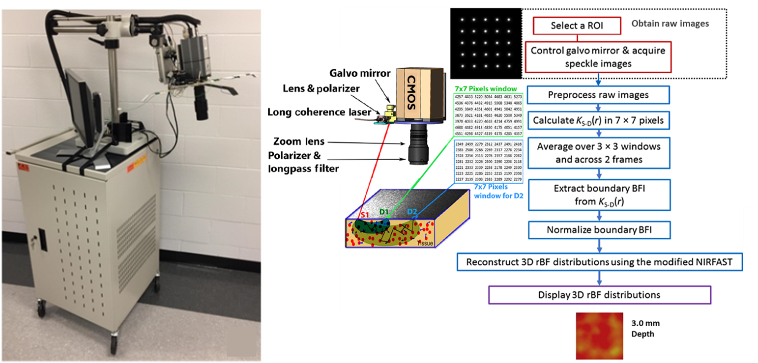
Speckle contrast diffuse correlation tomography (scDCT). Supported by the NIH, we have developed an innovative scDCT technology (US Patent #9861319), which provides a non-contact, fast, low-cost, and portable tool for high-density 2D/3D imaging of cerebral blood flow and oxygenation distributions in relatively deep brains (up to centimeter).
In case studies with the scDCT, continuous and longitudinal measurements of cerebral hemodynamics have been successfully demonstrated in brains of rodents, neonatal piglets, and human newborns. scDCT has also been used to image mastectomy skin flaps and burned/wound tissues.
Click image to view full screen
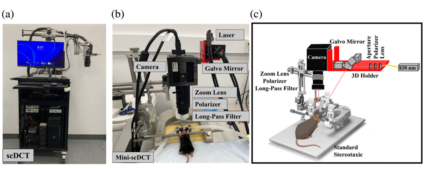
Fig. 1: Two scDCT systems. (a) A large and expensive scDCT system sitting on a movable cart. (b) A compact and affordable mini-scDCT sitting on a table. (c) Schematic of the mini-scDCT with optoelectronic components.
(Fatemeh Hamedi, Faezeh Akbari, Mehrana Mohtasebi, Chong Huang, Li Chen, Lei Chen, and Guoqiang Yu. “Affordable miniaturized speckle contrast diffuse correlation tomography device for depth-sensitive mapping of cerebral blood flow in rodents.” Journal of Biomedical Optics 30, no. 10 (2025): 106007-106007.)
Mini-scDCT
Leveraging a clinical scDCT system, we also developed an affordable, user-friendly, fast, and miniaturized scDCT (mini-scDCT) device tailored for depth-sensitive CBF imaging in small rodents.
The mini-scDCT replaces bulky and costly optoelectronic components with compact, low-cost alternatives while preserving imaging performance. It is mounted on a standard stereotaxic apparatus for portability and ease of use.
Temporal resolution was improved by hardware synchronization and software optimization. System validation was performed using head-simulating phantoms and rodent models under various pathophysiological conditions.
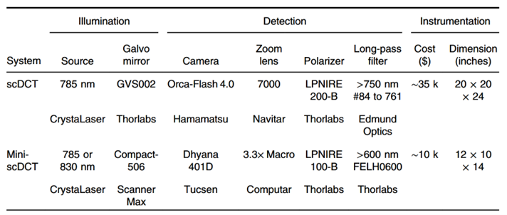
Table 1: Comparison between key parts/components for assembling the original scDCT and mini-scDCT devices.
Click image to view full screen

Click image to view full screen
Fig. 2: Depth-sensitive imaging of BFI maps from the head of a control mouse (Mouse #2, sham) at baseline. (a) Series of 2D BFI maps reconstructed using SDs ranging from 1.5 to 4 mm with BW =2 mm. Spatial resolution was quantified by selecting two ROIs on the 2D flow map (SD = 2 mm and BW = 2 mm): one over a large bundle of superior cerebral veins (L) and another over a small vessel(S). (b) Photograph of the mouse skull. (c) Corresponding height map of the skull. (d) Integrated 2D BFI map overlaid on the 3D head surface geometry.
(Fatemeh Hamedi, Faezeh Akbari, Mehrana Mohtasebi, Chong Huang, Li Chen, Lei Chen, and Guoqiang Yu. “Affordable miniaturized speckle contrast diffuse correlation tomography device for depth-sensitive mapping of cerebral blood flow in rodents.” Journal of Biomedical Optics 30, no. 10 (2025): 106007-106007.)
Click image to view full screen
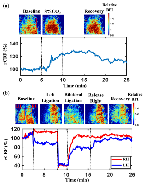
Fig. 3: Continuous monitoring of rCBF variations during 8% CO2 inhalation and transient carotid artery ligations was performed in a rat with the scalp retracted.
(a) BFI maps and corresponding time-course rCBF changes before, during, and after 8% CO2 inhalation. Dashed lines indicate the separation of experimental phases, including 5 min of baseline, 5 min of CO2 inhalation, and 15 min of recovery. (b) BFI maps and time-course rCBF changes before, during, and after sequential carotid artery ligations. Dashed lines delineate 3 min of baseline, 5 min of left carotid artery ligation, 2 min of bilateral ligation, 5 min following release of the right carotid artery, and 10 min of recovery after release of the left carotid artery. The temporal resolution was ∼2.5s.
(Fatemeh Hamedi, Faezeh Akbari, Mehrana Mohtasebi, Chong Huang, Li Chen, Lei Chen, and Guoqiang Yu. “Affordable miniaturized speckle contrast diffuse correlation tomography device for depth-sensitive mapping of cerebral blood flow in rodents.” Journal of Biomedical Optics 30, no. 10 (2025): 106007-106007.)